Bio Break: Understanding Continuous Glucose Monitors
Continuous glucose monitors (CGMs) are revolutionizing how people track blood sugar levels in real time. But how do they work, and where exactly do they measure glucose? In this episode of Bio Break, Nick and Joris explore the science behind CGMs, explaining the difference between blood glucose monitoring and interstitial fluid measurement—and why CGMs don’t actually draw blood continuously.
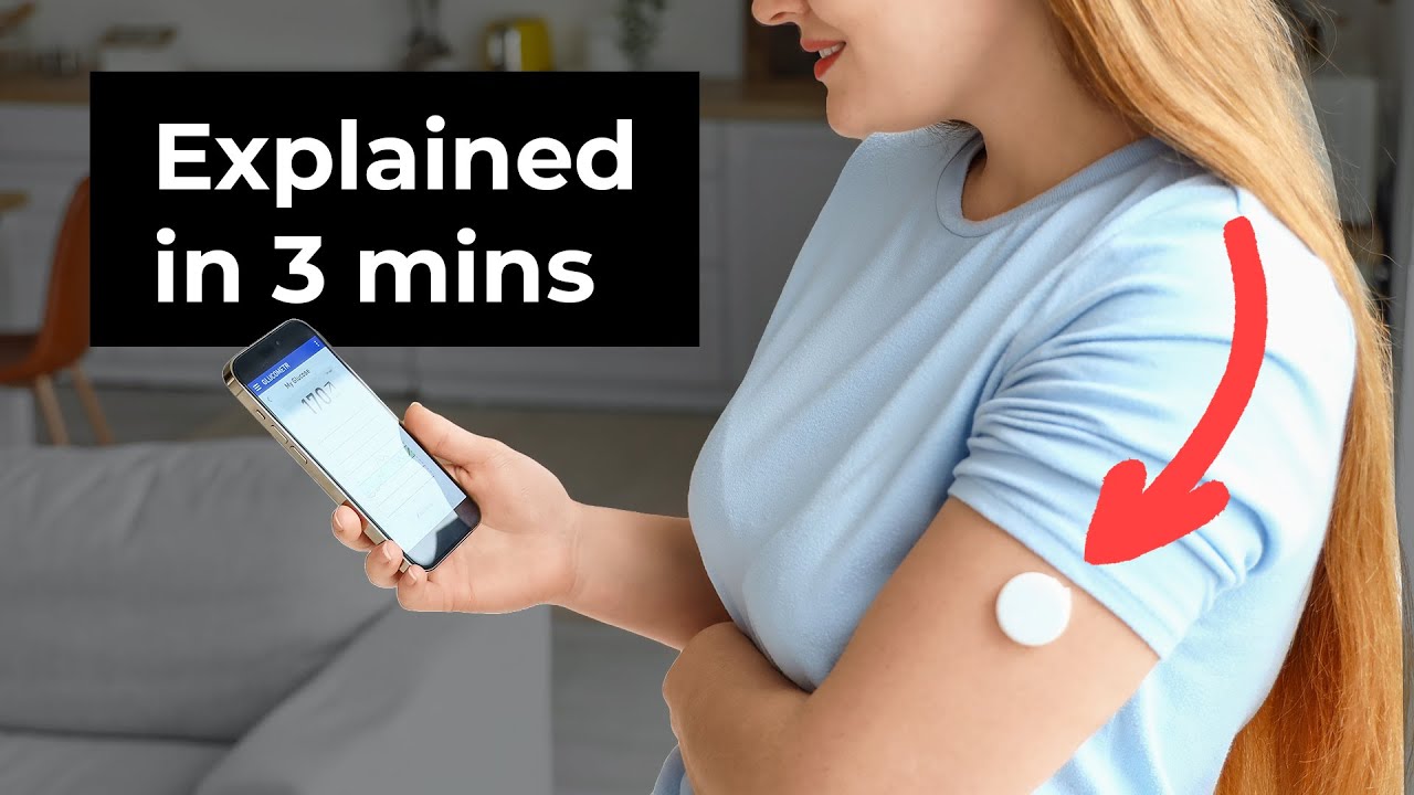
Rather than inserting a sensor directly into a vein, CGMs use microneedles or tiny tubes that sit in the interstitial fluid, the liquid surrounding the body’s cells. Unlike blood, which flows within veins, interstitial fluid is more accessible for continuous monitoring. However, there’s a key tradeoff—glucose levels in interstitial fluid lag behind blood glucose levels by about 5-10 minutes. This means CGMs don’t provide an instant blood sugar reading but rather a near-real-time estimate.
Nick raises an important question: does this time lag matter for diabetes management or sports performance tracking? Joris explains that while this delay exists, CGMs still offer huge advantages over traditional blood testing, which often requires finger pricks or lab draws that provide only a single data point. The ability to continuously measure glucose, lactate, or other small molecules gives users a clearer picture of how their body responds to food, exercise, and lifestyle changes.
However, not everything can be measured using interstitial fluid. Large molecules like hormones, red blood cells, and protein-bound compounds don’t diffuse as easily into interstitial fluid, making them difficult to track with a CGM. But for small molecules such as glucose, lactate, histamine, and creatinine, interstitial fluid monitoring is a powerful tool for real-time insights into metabolism.
If you’ve ever wondered how CGMs work or why they don’t require direct blood sampling, this episode breaks down the technology in a simple, engaging way.
Understanding Continuous Glucose Monitors
Related Resources

Nick and Nigel breaks down what actually goes into the cost of getting a sterilized device into a user’s hands, and why up to 30% of costs can sit in places most teams don’t plan for.
Theranostics combines diagnosis and therapy into a single targeting system, using one ligand to attach to two different radioactive payloads, one for imaging and one for treatment. It represents a significant shift in how cancer is being identified and treated. But the theranostics delivery workflow tells a different story.
Most medical devices were designed for clinical settings, not the patients and caregivers who increasingly rely on them at home. Here’s what good home-use device design actually requires.

How do you measure comfort in medical device design? Explore the tools, scales, and study design principles that turn a subjective experience into actionable design data.