Bio Break: Tooth-In-Eye Surgery
In this episode of Bio Break, Nick Allan and Joris van der Heijden dive into one of the most astonishing medical innovations we’ve ever come across: osteo-odonto-keratoprosthesis. Or, as Nick quickly dubs it, “tooth in eye surgery.”
This fascinating procedure sounds like science fiction but has been successfully used to restore vision in people who are blind due to damage in the front part of the eye, such as from trauma or autoimmune diseases. While the retina remains functional, traditional options are off the table. That’s where this extreme innovation comes in.
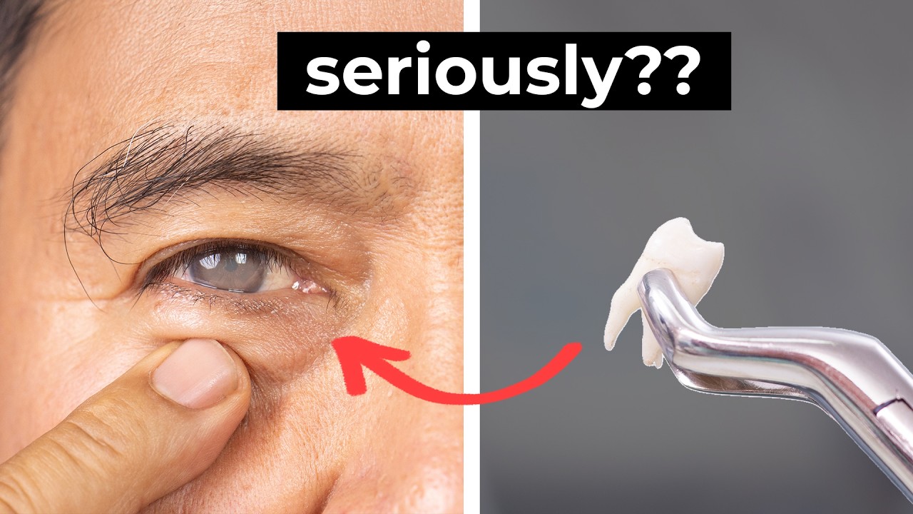
Joris explains how the procedure starts with removing a patient’s tooth and some surrounding bone to create a small square-shaped structure. A hole is drilled in the center, and a tiny lens is implanted, this piece will eventually act as an artificial cornea. But before it’s implanted into the eye, it needs to be biologically prepared. That’s done by temporarily placing the implant into the patient’s cheek, where it can become vascularized over a few months.
Once ready, the patient returns to the hospital for the second stage. Surgeons retrieve the now-living implant from the cheek and carefully insert it into the eye, replacing the damaged corneal area. Thanks to the previously grafted oral tissue, the eye is prepped to accept the implant, and the result is stunning: restored vision in up to 90% of patients.
Even more impressive? Around 50% of these individuals gain high-quality vision, enough to read or even consider driving.
Whether you’re a medtech enthusiast or just love mind-blowing medical stories, this episode is a must-watch. Learn how combining dental tissue, ocular surgery, and a bit of clever biology can give people their sight back.
Five Eye Diseases linked to majority of blindness or poor vision.
Related Resources
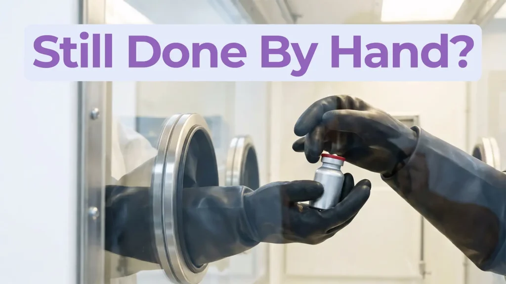
Theranostics combines diagnosis and therapy into a single targeting system, using one ligand to attach to two different radioactive payloads, one for imaging and one for treatment. It represents a significant shift in how cancer is being identified and treated. But the theranostics delivery workflow tells a different story.
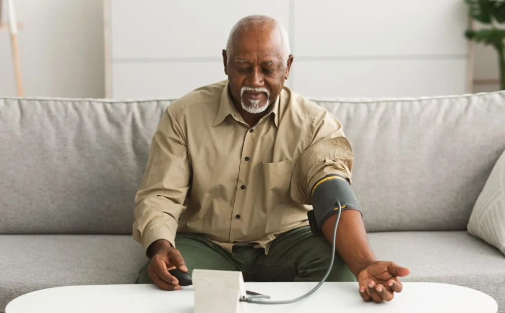
Most medical devices were designed for clinical settings, not the patients and caregivers who increasingly rely on them at home. Here’s what good home-use device design actually requires.
How do you measure comfort in medical device design? Explore the tools, scales, and study design principles that turn a subjective experience into actionable design data.
Gathering health data has enormous value for spotting risks, improving care, and advancing science. The problem isn’t capturing the data. The problem is how we choose to present it and who we’re really serving when we do.