
ELISA Assay Explained Using Simple Models
Understanding how diagnostic assays work is critical for anyone developing or evaluating medical tests. In this Bio Break episode, Nick and Nigel break down the ELISA assay explained in simple, practical terms using everyday models. Rather than relying on dense theory, they walk through how antibodies, antigens, and enzymes work together to create a detectable signal. This clear explanation helps demystify a tool that is widely used in diagnostic development and laboratory testing.
What Is an ELISA Assay
An ELISA assay, short for enzyme linked immunosorbent assay, is a common laboratory method used to detect specific biological targets. These targets are often antigens, which can include proteins associated with disease or infection. In this episode, Nick explains that ELISA assays rely on antibodies that are designed to bind to a specific target with high precision.
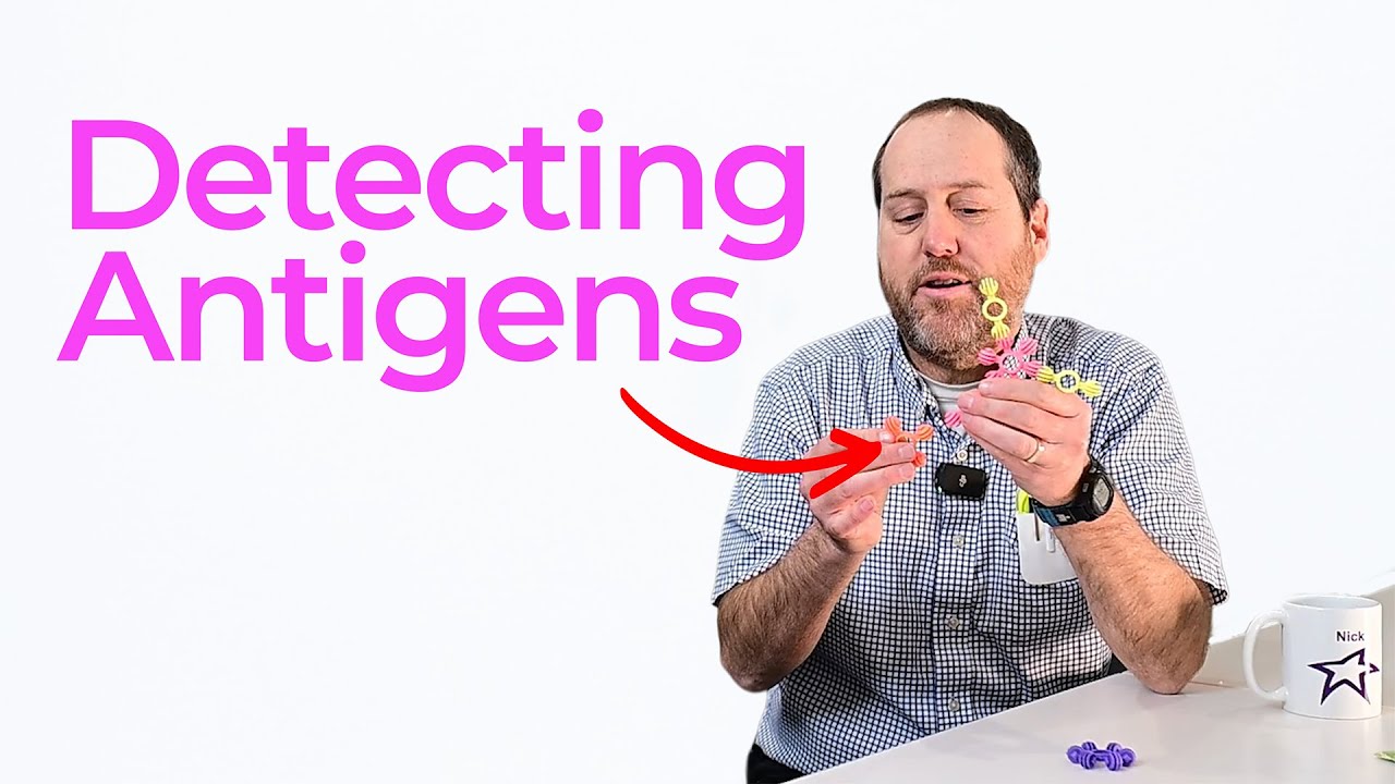
To make the concept approachable, Nick uses toy models to represent monoclonal antibodies. These antibodies all come from a single immune cell, which means they are identical. As a result, they consistently bind to the same antigen. This consistency is one of the reasons ELISA remains a trusted diagnostic tool.
ELISA Assay Explained Through Antibodies and Antigens
When looking at an ELISA assay explained step by step, antibodies are the key detection component. Each antibody has a conserved region and a variable region. The variable region is responsible for recognizing the antigen, while the conserved region signals that the antibody belongs to the immune system.
In the example shown, antigens are placed into a well plate. Antibodies that have been linked to an enzyme are then introduced. If the antigen is present, the antibody binds to it. This binding event is the foundation of detection in an ELISA assay.
Direct ELISA and Signal Detection
This Bio Break focuses on the direct ELISA format. In a direct ELISA, the antibody is directly linked to an enzyme. Once binding occurs, a substrate is added. The enzyme reacts with the substrate and produces a visible color change. In this case, the signal turns green, confirming that the antigen has been detected.
Although Nick notes that this is one of the simplest ELISA formats, it clearly demonstrates the core principle. Detection happens because biology and chemistry are intentionally paired to create a readable signal.
Why ELISA Matters in Diagnostic Development
ELISA assays are widely used in diagnostic development, including applications such as laboratory testing and rapid diagnostic concepts. By understanding how ELISA works at a fundamental level, teams can make better decisions about assay design, limitations, and future development paths. This episode sets the stage for deeper discussions on more advanced detection formats.
This Bio Break episode shows that complex diagnostic tools do not have to be intimidating. By walking through an ELISA assay explained with clear language and physical models, Nick and Nigel make the science accessible while staying grounded in real laboratory practice.
Enjoying Bio Break? Sign up to get new episodes sent to your inbox.
Related Resources
Many teams still underuse CM&S, often bringing it late in device validation, when key decisions have already been made. That approach leaves much of the value of CM&S untapped.

This article traces the Pennes bioheat equation from its 1948 origins to modern multiscale approaches, explaining how engineers select the right level of modelling complexity across device categories.

Five methods for building accurate project estimates, from gut feel to bottom-up, plus three add-ons that sharpen any estimate.

Designers and engineers today have no shortage of tools to improve designs, accelerate timelines, and meet cost targets. The challenge is knowing what to use and when.