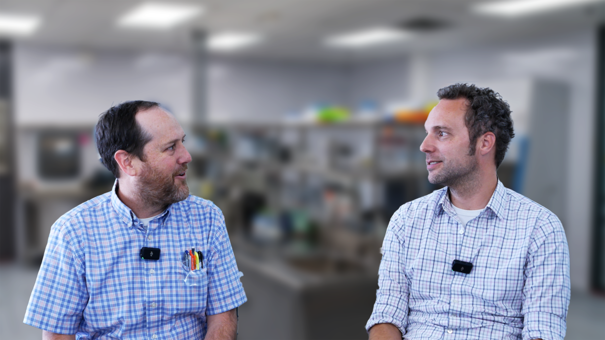
Bio Break: Real-Time Imaging for Targeted Drug Delivery
In this episode of Bio Break, Nick and Joris discuss the fascinating world of real-time imaging for targeted drug delivery. When delivering drugs to precise locations in the body, how do we ensure they reach the right spot? The answer lies in medical imaging technologies such as MRI, CT, and ultrasound, which play a crucial role in guiding complex drug delivery devices.
Nick starts by asking how imaging technologies help visualize drug targeting during delivery. Joris explains that MRI (Magnetic Resonance Imaging), CT (Computed Tomography), and ultrasound are the three most commonly used imaging techniques. Each has its advantages and limitations depending on the application.
- MRI offers exceptional resolution and is ideal for deep-tissue targeting, such as brain delivery for conditions like Parkinson’s disease. However, intraoperative MRI machines are less accessible in hospitals, making their real-time use challenging.
- CT scanning is more widely available and excellent for bone-related targeting, such as spinal drug delivery. While it has slightly lower resolution than MRI, it is a practical and widely adopted solution in medical settings.
- Ultrasound is the most real-time imaging method and is commonly used for nerve blocks and pain management. However, it has depth limitations and is ineffective for regions behind bone, such as the brain.
Nick and Joris also discuss the future of medical imaging in drug delivery. With advancements in 3D imaging and computational modeling, physicians will soon have better spatial awareness of the drug delivery process, improving precision and patient outcomes.
As medical imaging evolves, AI-driven technologies and 3D imaging integration will revolutionize precision drug delivery, making it more efficient and accessible.
Real-Time Imaging for Targeted Drug Delivery
Related Resources

Nigel Syrotuck breaks down REACH SVHC compliance for teams working with material suppliers and compliance questionnaires.

We examine when computational modelling and simulation, or CM&S, genuinely supports medical device simulation strategy and when it becomes a costly detour.
Many teams still underuse CM&S, often bringing it late in device validation, when key decisions have already been made. That approach leaves much of the value of CM&S untapped.

This article traces the Pennes bioheat equation from its 1948 origins to modern multiscale approaches, explaining how engineers select the right level of modelling complexity across device categories.