
Convection-Enhanced Delivery: Infusing Precision into Neurosurgery
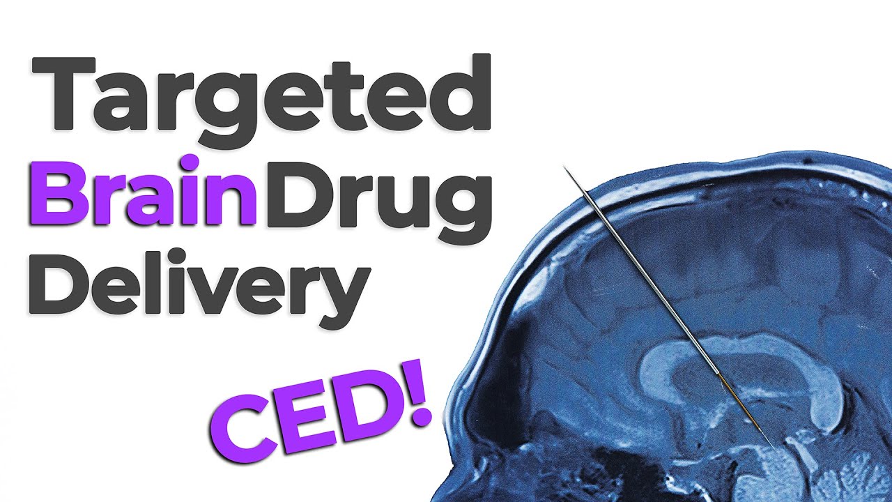
In this episode of MedDevice by Design, we explore how convection-enhanced delivery (CED) is transforming brain-targeted drug therapies. Ariana Wilson and Mark Drlik explain how this specialized method delivers therapeutic agents directly into brain tissue using precision-guided devices for safe and accurate infusion.
What Is Convection-Enhanced Delivery?
Convection-enhanced delivery is a neurosurgical technique that infuses medication directly into the brain. Unlike passive diffusion, CED uses pressure gradients to push molecules more uniformly and deeply into brain tissue. This approach enables better distribution of therapeutic agents to target areas.
Cannula Design
CED requires a rigid yet precise neurosurgical cannula. Surgeons place the cannula using a neuro-navigation system and preoperative imaging, which help chart the safest path into the brain. With over 600 kilometers of vasculature—equivalent to stacking 2,000 Eiffel Towers—avoiding blood vessels is critical.
Once placed, some cannulas become flexible to support long-term infusion and reduce infection risk by routing the tubing away from the wound site.
Navigating the Brain with Surgical Precision
Placing a cannula into the brain is a complex process. Neurosurgeons depend on real-time imaging and navigation tools to avoid sensitive areas. This level of surgical precision is what makes CED so promising for targeted brain therapies.
Enjoying MedDevice by Design? Sign up to get new episodes sent to your inbox.
Related Resources
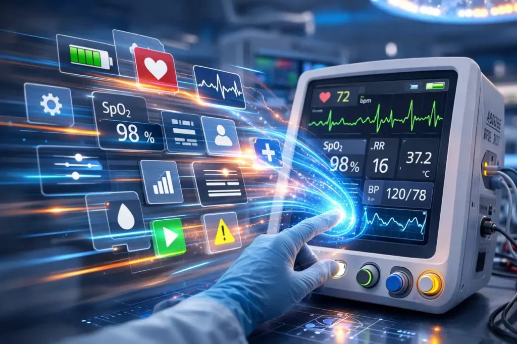
Medical device teams developing embedded and cross-platform GUIs can accelerate delivery without compromising usability or validation by choosing the right framework early and designing for performance, portability, and maintainability.
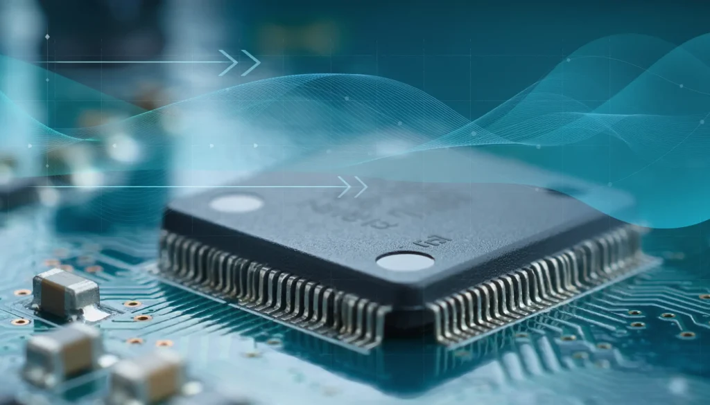
Compute demands on “the edge”, like embedded sensors or remote devices. have grown significantly as AI has moved from experimentation to deployment. Medical devices are pushing more of their AI functionality onto edge hardware.
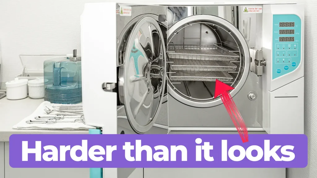
Medical device cleaning is more complex than it seems. In this Bio Break episode, Nick and Nigel unpack what really goes into cleaning medical devices and why it cannot be treated like a simple wipe-down process.
This blog reviews the main families of optical detectors and the major technologies in those families.