
Understanding Contrast Agents in Imaging
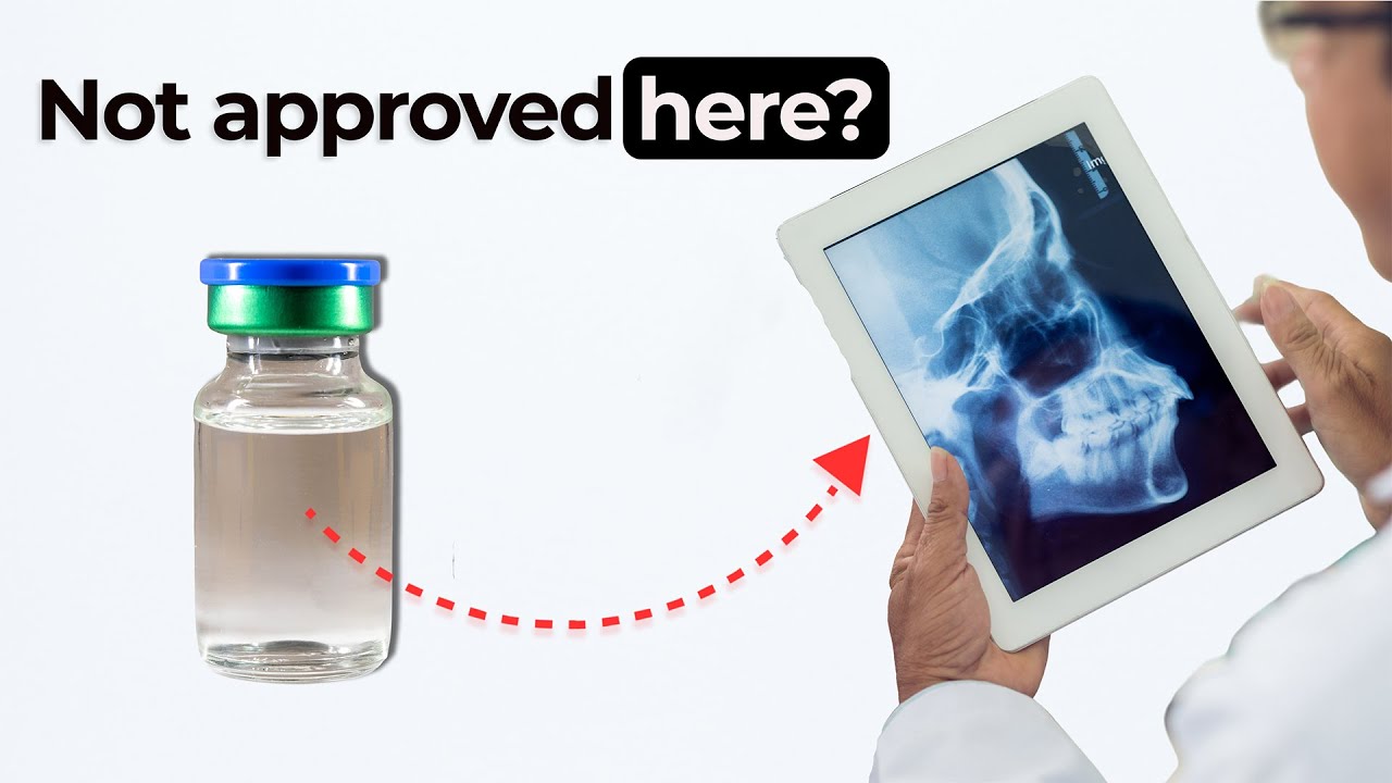
In this episode of Bio Break, Nigel and Nick explore how contrast agents in imaging support medical device trials and diagnostics. While bones appear clearly in standard X-rays, soft tissues like those in the nasal cavity often require contrast agents to become visible. These agents enhance the effectiveness of imaging by highlighting specific anatomical features.
Nick shares how contrast is key to delivering drugs precisely, while Nigel explains the challenges of finding approved contrast agents for delicate regions like the nasal passages. They describe trials involving soft tissue imaging and how regulatory concerns can slow adoption. The issue isn’t always about safety or performance—it’s about risk mitigation. If a contrast agent lacks a predicate or history of use in a specific body region, developers face extra hurdles to prove it’s safe and effective.
Barium salts come up as a classic example of a commonly approved agent, although not all are pleasant to work with. The episode also reflects on how these small details—like tissue compatibility or long-term absorption—impact trial design and device approval.
As always, Nigel and Nick keep it light while highlighting the complexities engineers face when designing trials that involve advanced imaging and anatomical targeting.
Want more on material testing or clinical trials? Watch our episode on drop testing medical devices or learn how sterilization methods impact device usability and safety.
Related Resources

Understanding gram positive vs negative bacteria is essential when studying sterility, microbiology, and antibiotic effectiveness. While many people think the difference is only about staining, the reality is much deeper.
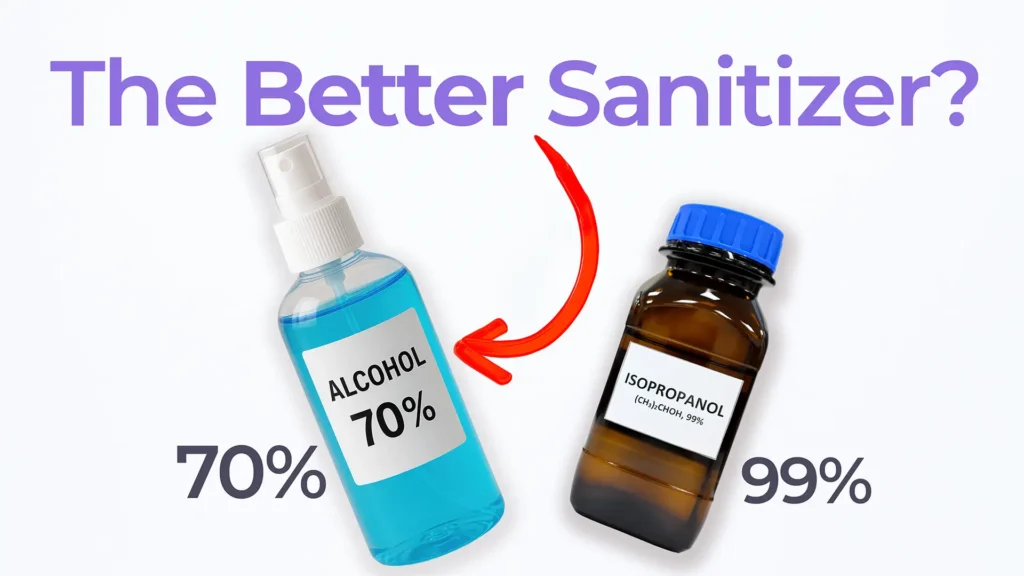
Nick and Nigel explore the science behind hand sanitizer formulations. They discuss how alcohol interacts with bacterial cells, why water improves its effectiveness, and what the additional ingredients in sanitizer actually do.

Antibiotics changed medicine forever, but many people still wonder how penicillin works at a biological level. Nick and Nigel break down the science behind one of the most important antibiotics ever discovered.
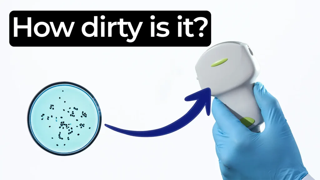
Nick and Nigel explore how much bacteria can exist on devices and why it matters. They explain that bacteria are everywhere.